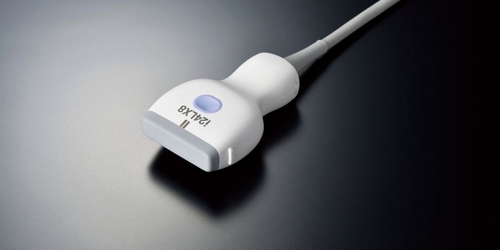
High-frequency ultrasound probes deliver improved visualisation of bones, joints & soft tissues for patients at Addenbrooke’s Hospital
The widening range of diagnostic ultrasound probes available on the Aplio i-series ultrasound system is expanding the possibilities for Musculoskeletal (MSK) radiology clinical practice and reducing the need for MRI scan appointments at Addenbrooke’s Hospital, part of Cambridge University Hospitals NHS Foundation Trust. Using high-frequency 24 MHz and 33 MHz linear matrix technology probes, the clinical team is gaining improved anatomical detail of superficial structures such as nerves and tendons in hands and fingers. This will help with hand and wrist injury explorations and provide greater detail in pre and post-operative examinations.
“The advantages of the innovation in high-frequency ultrasound probes mean gaining greater imaging detail to support clinical confidence. This includes giving excellent image resolution of superficial or close-to-skin surface structures such as nerves under the skin, to help identify damage following surgery or explore causes of a patient’s loss of sensation,” states Dr Andrew Grainger, Consultant MSK Radiologist at Cambridge University Hospitals NHS Foundation Trust.
The improved image quality from new probes improve capabilities to explore tendon injuries in fingers and hands. This can be to assess subtle damage from glass or knife injuries or post-operative issues.
There are also time and process savings that the development in ultrasound probes will also help with. For example, it takes 15 minutes to do a complicated MSK ultrasound scan compared to 30-45 minutes for an MRI scan and the additional administration associated with the paperwork for patient consent. So, being able to do a simple ultrasound examination and gain superb image detail to base decisions on is much better for improving the patient experience and care journey. Furthermore, it is this improvement in image quality that will also help with surgical planning. Traditionally, surgeons like MRI scan images as they show anatomical detail as true-to-life for procedural planning. With ultrasound, clinicians are now able to deliver comparable or even better superficial imaging, there is the potential to change the way radiology and surgery work together.
Iain Dunn, Business Development Manager & Territory Sales Manager Ultrasound at Canon Medical Systems UK states, “The constant development of medical technology is adding more advantages to clinical practice efficiency and improving the quality of patient care. The evolution in high-frequency ultrasound transducers means that it isn’t only the system that needs to be selected, but also the probes that are designed to broaden clinical application and offer better imaging outcomes compared to lower frequency probes and some MRIs. It is great to hear that patients at Addenbrooke’s Hospital are already benefitting from the innovation and that their MSK care is being enhanced.”


